کتاب مصور انگل شناسی با عنوان graphic images of parasites یک اطلس رنگی انگل شناسی است که بسیاری از انگل های انسانی و حیوانی و بند پایان مختلف را به صورت کاملاً تصویری و به ترتیب حروف الفبا، نمایش می دهد.
در این کتاب در مورد هر انگل توضیحاتی در خصوص معرفی کلی، مورفولوژی، سیر تکاملی و بیماری زایی آن توضیح داده می شود.
این کتاب در ۳۴۴ صفحه با فرمت pdf و به زبان انگلیسی می باشد.
در ادامه لینک دانلود و بخش هایی از تصاویر و متون این کتاب قابل مشاهده می باشد.
جهت دانلود کتاب مصور انگل شناسی کلیک کنید:
صفحاتی از کتاب مصور انگل شناسی
مبحث سوسک آمریکایی

Periplaneta americana
(American cockroach)
Cockroaches can disseminate a number of human parasitic diseases. For example, their external surfaces could be contaminated with protozoan cysts or helminth eggs, and these infectious stages could be deposited in areas where one would not expect to find the cysts or eggs. “Filth flies” can disseminate infections by the same mechanism. In this sense, cockroaches serve as mechanical vectors. Cockroaches do not, however, serve as biological vectors for any parasitic diseases of humans.
The American cockroach, Periplaneta americana, is the intermediate host for the acanthocephalan, Moniliformis moniliformis. This is a parasite of the small intestines of rats, and it has been used as an experimental model to study the biology of acanthocephala.
مبحث آنکیلوستوما

Ancylostoma spp. and Necator spp.
(hookworms)
There are many species of hookworms that infect mammals. The most important, at least from the human standpoint, are the human hookworms, Ancylostoma duodenale and Necator americanus, which infect an estimated 800,000,000 persons, and the dog and cat hookworms, A. caninum and A. braziliense, respectively. Hookworms average about 10 mm in length and live in the small intestine of the host. The males and females mate, and the female produces eggs that are passed in the feces. Depending on the species, female hookworms can produce 10,000-25,000 eggs per day.
About two days after passage the hookworm egg hatches, and the juvenile worm (or larva) develops into an infective stage in about five days. The next host is infected when an infective larva penetrates the host’s skin. The juvenile worm migrates through the host’s body and finally ends up in the host’s small intestine where it grows to sexual maturity. The presence of hookworms can be demonstrated by finding the characteristic eggs in the feces; the eggs can not, however, be differentiated to species (view diagram of the life cycle).
The mouthparts of hookworms are modified into cutting plates. Attachment of hookworms to the host’s small intestine causes hemorrhages, and the hookworms feed on the host’s blood. Hookworm disease can have devastating effects on humans, particularly children, due to the loss of excessive amounts of blood.
Juveniles (larvae) of the dog and cat hookworms can infect humans, but the juvenile worms will not mature into adult worms. Rather, the juveniles remain in the skin where they continue to migrate for weeks (or even months in some instances). This results in a condition known as “cutaneous” or “dermal larval migrans” or “creeping eruption.” Hence the importance of not allowing dogs and cats to defecate indiscriminately.
مبحث سیکلوسپورا

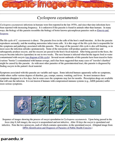
Cyclospora cayetanensis
Cyclospora cayetanensis infections in humans were first reported in the late 1970’s, and since that time infections have been reported with increasing frequency. It is unknown if this parasite is found in animals other than humans. In many ways, the biology of this parasite resembles the biology of better known apicomplexan parasites such as Eimeria and Isospora.
The life cycle of C. cayetanensis is direct. The parasite lives in the cells of the host’s small intestine. At first the parasite reproduces asexually, and the resulting merozoites infect more cells. It is this stage of the life cycle that is responsible for the symptoms and pathology associated with this parasite. This stage of the parasite’s life cycle is also self-limiting, so in most cases the infection subsides spontaneously.
Some of the merozoites will produce gametes which fuse and subsequently produce oocysts, and the oocysts are passed in the host’s fecal material. After being passed in the feces, the oocysts become infective (sporulate) in one to two weeks. The next human is infected when he/she ingests food or water contaminated with oocysts (see diagram of life cycle).
Several recent outbreaks of this parasite have been traced to fruits (various “berries”) contaminated with human sewage, and it has been suggested that many cases of “traveler’s diarrhea” might be caused by this parasite. As with most other parasites of the gastrointestinal tract, this parasite is diagnosed by finding oocysts in the patient’s fecal material.
Symptoms associated with this parasite are variable and vague. Some infected humans apparently suffer no symptoms, while others suffer various degrees of diarrhea, gas, cramps, nausea, vomiting, and fever. In most instances these symptoms disappear in a few days, but in some cases the symptoms may last for months. Prescription drugs are available that will cure the infection. It is not known if humans with compromised immune systems (e.g., AIDS patients) suffer more serious symptoms.
کتاب مصور انگل شناسی قابل استفاده برای اساتید و تمام دانشجویان رشته های مختلف دانشگاهی می باشد.
 انگل شناسی پزشکی Medical Parasitology


